CAI researchers develop advanced AI model for medical image processing in full 3D
A new publication from the Intelligent Vision Systems group (IVS) at CAI introduces an AI method to reduce patient radiation dose in cancer therapy.
Cone-beam Computed Tomography (CBCT) is frequently used on-board of clinical radiation therapy devices for patient positioning and tumor targeting. However, the clinical necessity of reducing patient radiation dose due to the imaging process via sparse-view sampling renders conventional reconstruction methods ineffective.
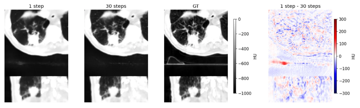
In a recent publication in the journal IEEE Access, the team around Daniel Barco, Dr. Marc Stadelmann and Dr. Frank-Peter Schilling of CAI’s Intelligent Vision Systems (IVS) group introduced MInDI-3D, a novel iterative diffusion-based model which is able to augment medical images in full 3D, and applied it to sparse-view CBCT images.
Addressing the challenge of sparse-view CBCT reconstruction was the goal of the international research project AC3T funded by Innosuisse, bringing together two ZHAW institutes, CAI and the Institute of Applied Mathematics and Physics (IAMP), Varian Medical Systems, part of Siemens Healthineers and world market leader in clinical radiation therapy devices, as well as collaborators from Yonsei University and Hospital, Seoul, South Korea.
MInDI-3D combines a U-Net based model with attention mechanisms and uses an iterative process to efficiently refine a sparse-view CBCT volume. Unlike traditional methods, MInDI-3D explicitly navigates the perception-distortion trade-off, allowing for high-fidelity 3D reconstructions even from sparse data. To ensure robust training, a novel large-scale pseudo-CBCT dataset of more than 16000 scans was produced and made publicly available.
The model’s performance was comprehensively evaluated using quantitative metrics, task-based assessments involving automated organ segmentation, and a qualitative review by a panel of clinicians at Yonsei University Hospital in South Korea. MInDI-3D demonstrated excellent performance in removing artifacts and restoring image quality. Furthermore, MInDI-3D generalized successfully to different scanner hardware, and the clinical assessment yielded favourable results.
MInDI-3D successfully addresses the critical clinical need to lower radiation dose without compromising image quality. The strong quantitative results, robust generalization, and evaluation by clinicians underscore its potential as a practical tool for clinical workflows, particularly in adaptive radiation therapy.