Automated Measurement of Tissue Velocities in Echocardiography
In a collaboration with the Zürich Startup MediRapp AG, we trained a computer vision model to automatically detect clinically relevant peak velocities in echocardiographic tissue Doppler traces. The vendor-agnostic approach harmonizes measurements across sites, improves consistency, comparability, and longitudinal tracking without device-specific calibration or tooling.
In this study, we present a vendor-agnostic,
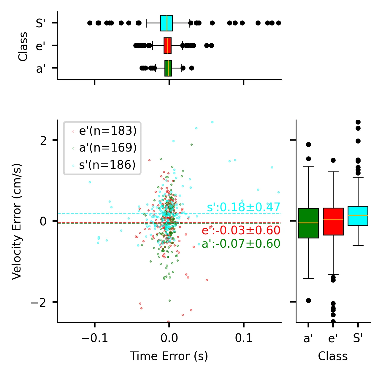
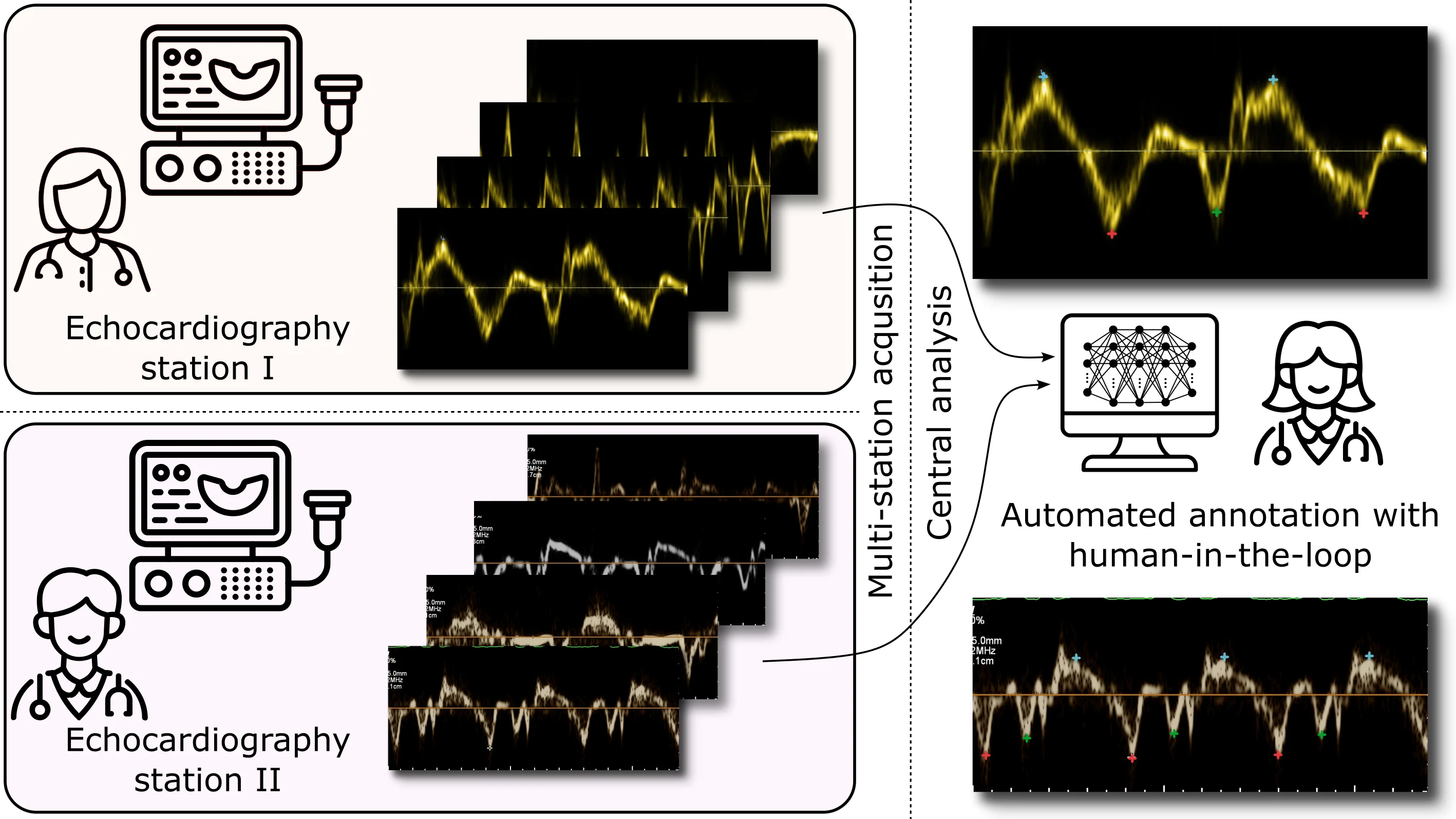
deep learning-based system for the automated analysis of transthoracic pulsed-wave tissue Doppler imaging (TDI),which decouples image acquisition from interpretation and enables centralized, fleet-wide analysis across devices. The model ingests standard TDI from heterogeneous ultrasound systems and automatically extracts key diagnostic markers peak systolic velocity (S′), early diastolic velocity (e′), and late diastolic/atrial contraction velocity (a′) using a single, unified pipeline. Conceptually, this harmonizes measurements across vendors and sites, improving consistency, comparability, and longitudinal tracking without device-specific calibration or tooling. Procedurally, a central inference service supports asynchronous batch processing and human-in-the-loop review, thereby shifting analysis off-console, allowing ultrasound scanners to remain fully available for acquisition. In our clinical dataset, which spans two ultrasound vendors and diverse cardiac cycles, the system correctly identified more than 93% of tissue velocity landmarks. In 50% of studies, all automated detections matched expert annotations, eliminating the need for manual edits. This approach streamlines offline TDI analysis, accelerates turnaround, and supports scalable, standardized cardiac assessments.
Full preprint: Automated, vendor-agnostic measurement of myocardial tissue velocities in echocardiography
Infos about the project partner: MediRapp AG